| Catalog No. | HX250013 |
| Host species | Mouse |
| Species reactivity | Human |
| Form | Liquid |
| Storage buffer | 0.01M PBS, pH 7.4. |
| Purity | >95% as determined by SDS-PAGE. |
| Clonality | Monoclonal |
| Isotype | IgG2b-kappa |
| Applications | ELISA, FCM, WB |
| Target | Beta-2-microglobulin, Beta-2-microglobulin form pI 5.3, B2M, CDABP0092, HDCMA22P |
| Purification | Protein A/G purified from cell culture supernatant. |
| Endotoxin level | Please contact with the lab for this information. |
| Accession | P61769 |
| Stability and Storage | Use a manual defrost freezer and avoid repeated freeze-thaw cycles. Store at 4°C short term (1-2 weeks). Store at -20°C 12 months. Store at -80°C long term. |
| Clone ID | BBM.1 |
| Note | For research use only. |
Anti-Human B2M/Beta-2-microglobulin Antibody (BBM.1)
Overview
Images
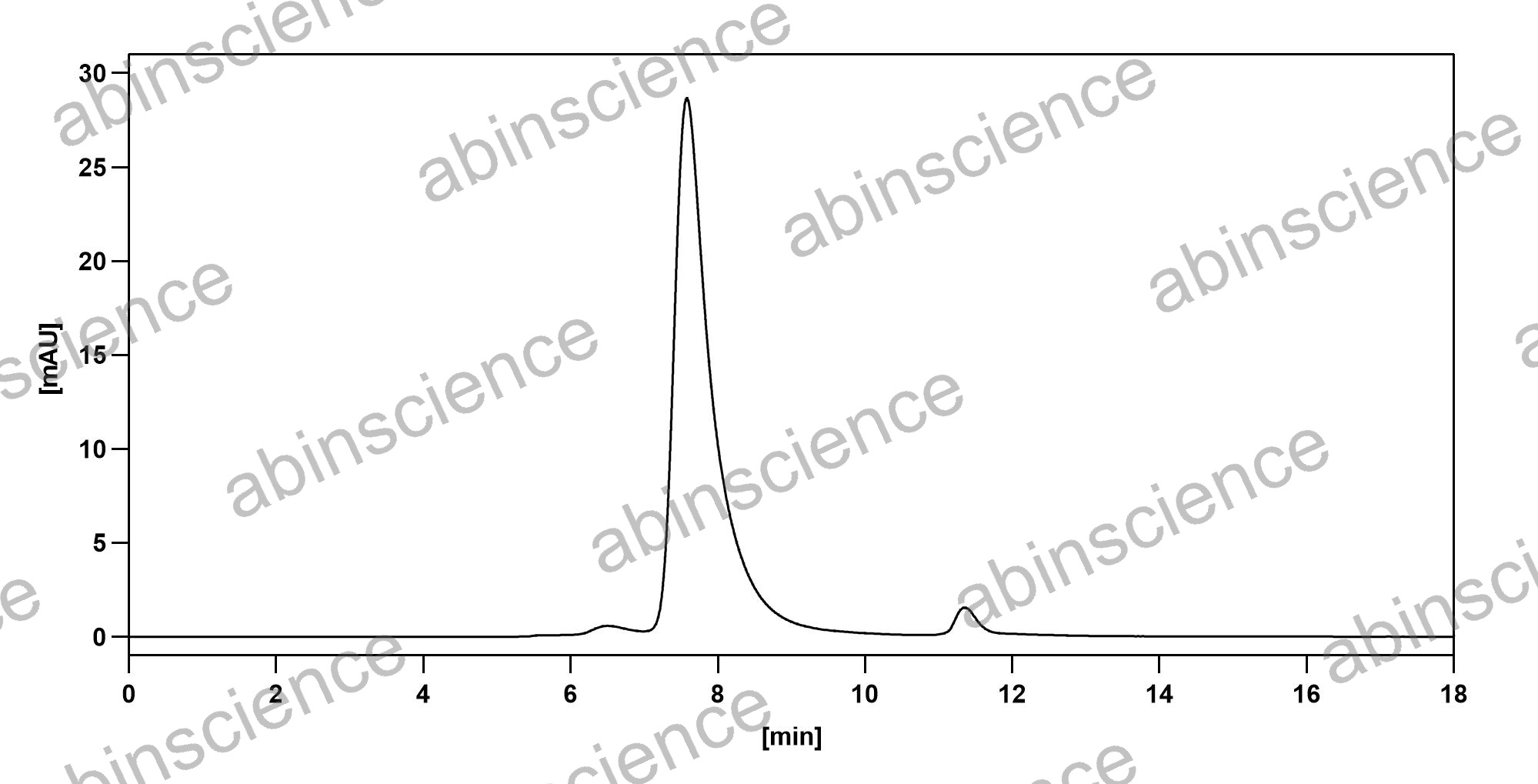 SEC-HPLC |
SEC-HPLC detection for Anti-Human B2M/Beta-2-microglobulin Antibody (BBM.1). |
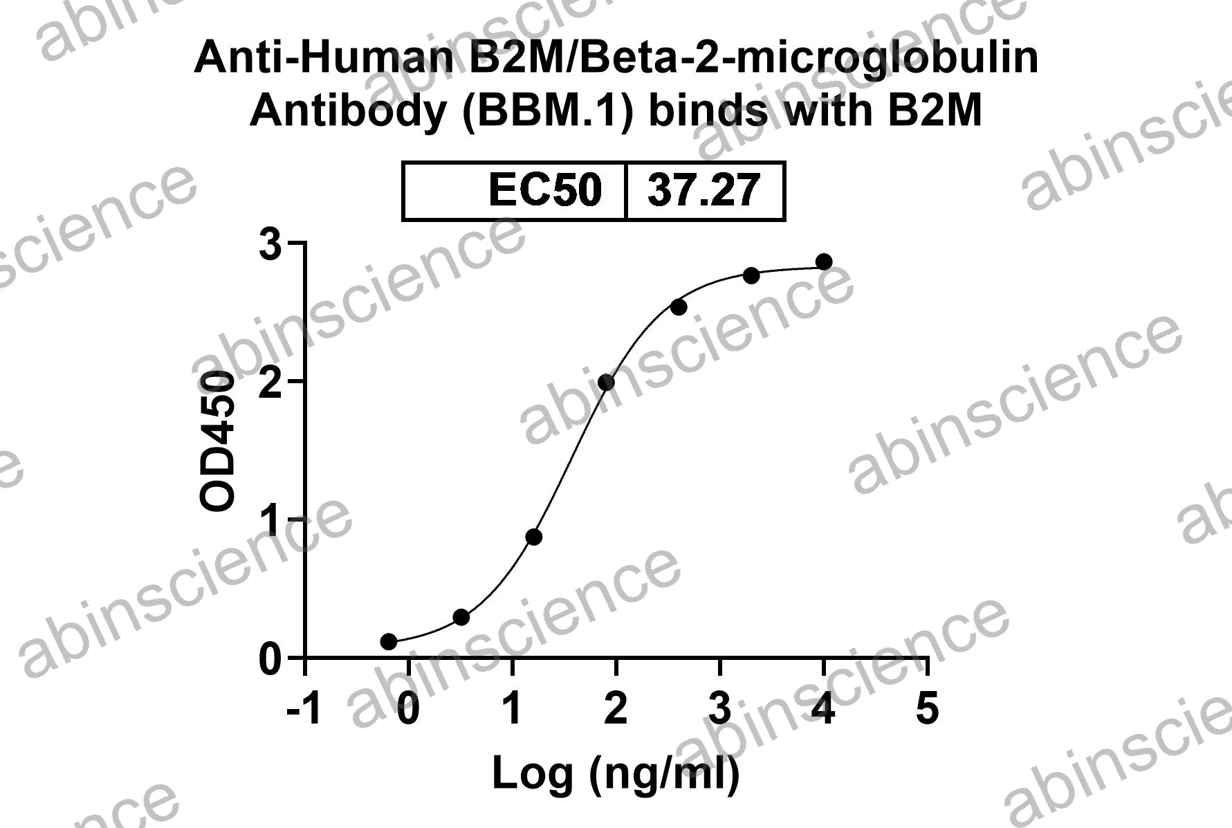 Bioactivity |
Detects B2M/Beta-2-microglobulin in indirect ELISAs. |
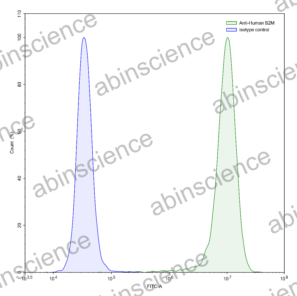
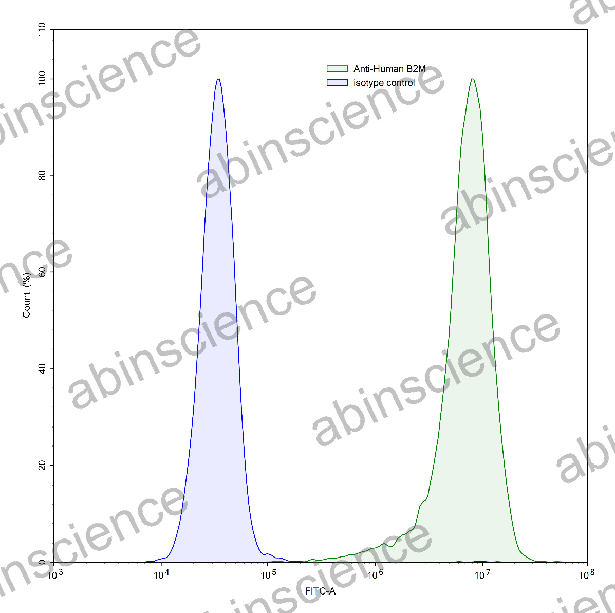 Flow Cytometry |
Flow-cytometry using anti-human B2M antibody.HeLa cells were stained with an irrelevant antibody (Blue Histogram) or an anti-human B2M antibody monoclonal antibody (Catalog # HX250013 ,Green Histogram) at a concentration of 5 µg/ml for 30 mins at RT. After washing, bound antibody was detected using a FITC conjugated goat anti-mouse antibody (Catalog # MF690414) and cells analysed on a NovoCyte Flow Cytometer. |
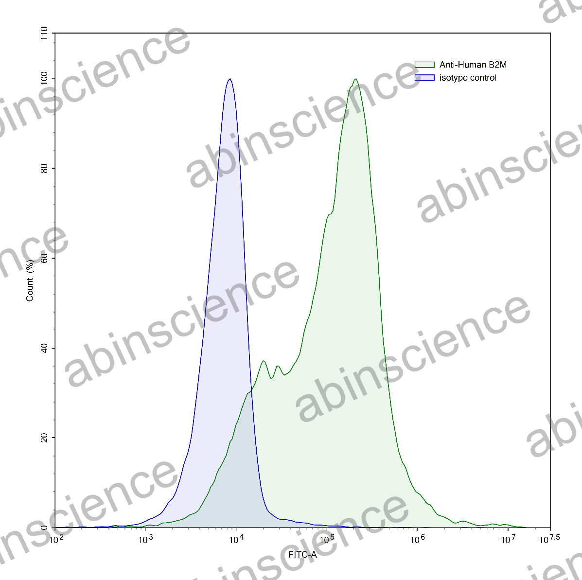
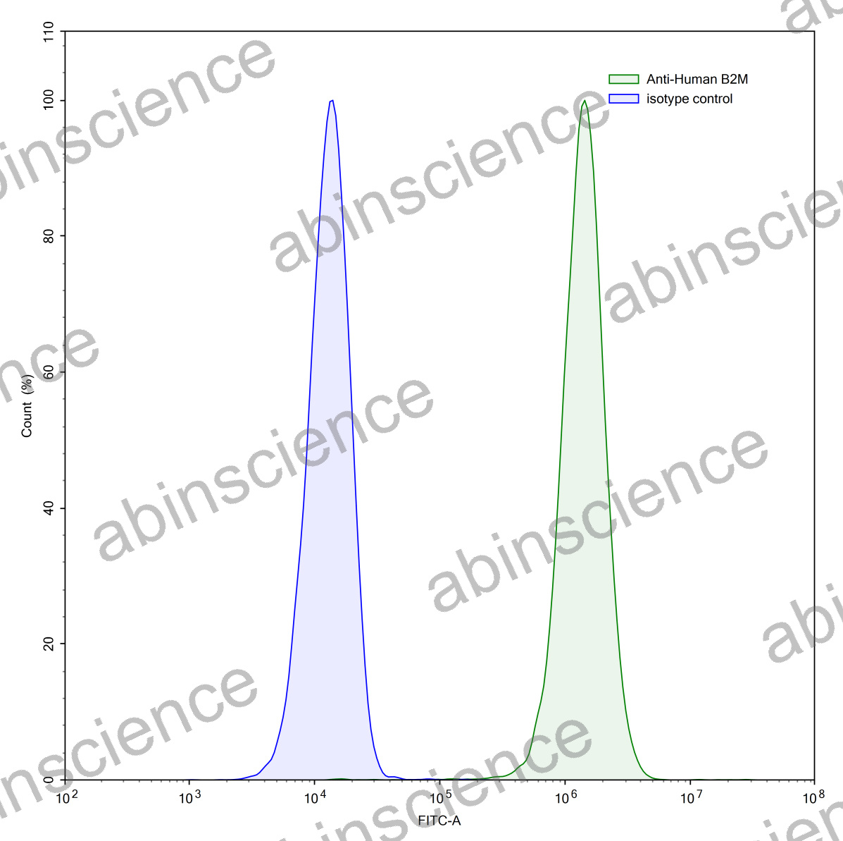 Flow Cytometry |
Flow-cytometry using anti-human B2M antibody.Jurkat cells were stained with an irrelevant antibody (Blue Histogram) or an anti-human B2M antibody monoclonal antibody (Catalog # HX250013 ,Green Histogram) at a concentration of 5 µg/ml for 30 mins at RT. After washing, bound antibody was detected using a FITC conjugated goat anti-mouse antibody (Catalog # MF690414) and cells analysed on a NovoCyte Flow Cytometer. |
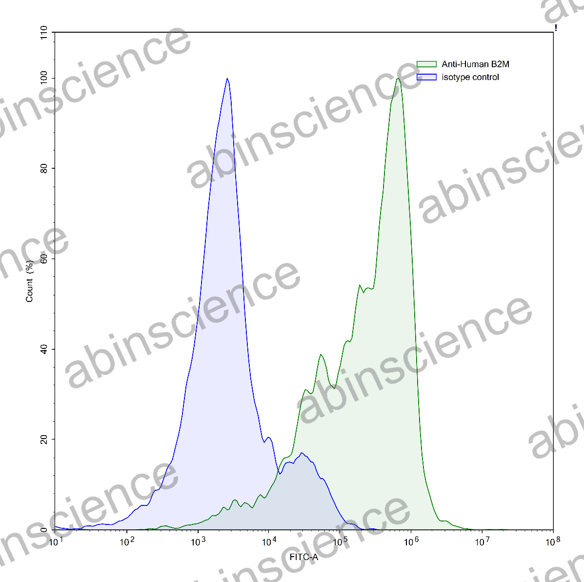
 Flow Cytometry |
Flow-cytometry using anti-human B2M antibody.A431 cells were stained with an irrelevant antibody (Blue Histogram) or an anti-human B2M antibody monoclonal antibody (Catalog # HX250013 ,Green Histogram) at a concentration of 5 µg/ml for 30 mins at RT. After washing, bound antibody was detected using a FITC conjugated goat anti-mouse antibody (Catalog # MF690414) and cells analysed on a NovoCyte Flow Cytometer. |
 Flow Cytometry |
Flow-cytometry using anti-human B2M antibody.Human peripheral blood lymphocytes were stained with an irrelevant antibody (Blue Histogram) or an anti-human B2M antibody monoclonal antibody (Catalog # HX250013 ,Green Histogram) at a concentration of 5 µg/ml for 30 mins at RT. After washing, bound antibody was detected using a FITC conjugated goat anti-mouse antibody (Catalog # MF690414) and cells analysed on a NovoCyte Flow Cytometer. |
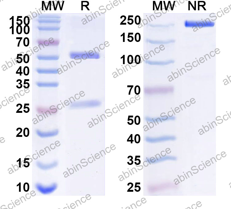 SDS-PAGE |
SDS-PAGE for Anti-Human B2M/Beta-2-microglobulin Antibody (BBM.1). |
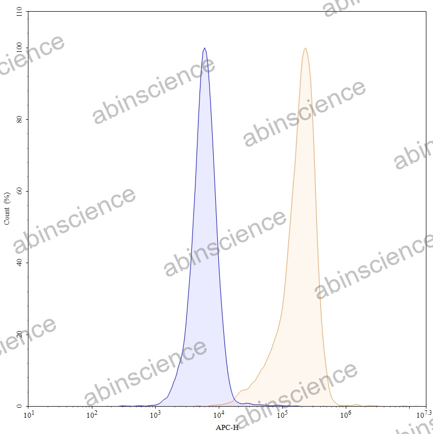 Flow CytoMetry |
Flow-cytometry using APC anti-human B2M antibody. Hela cells were stained with an irrelevant antibody (Blue Histogram) or an APC anti-human B2M monoclonal antibody (Catalog: HX250013, Yellow Histogram) at a concentration of 5 µg/ml for 30 mins at RT. After washing, and cells analysed on a NovoCyte Flow Cytometer. |
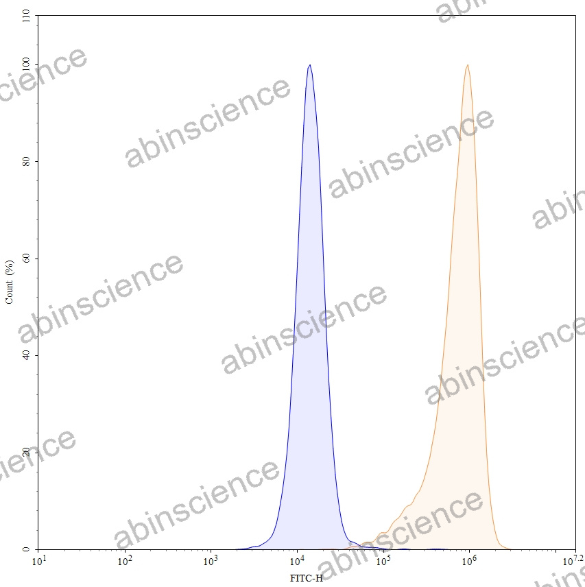 Flow CytoMetry |
Flow-cytometry using FITC anti-human B2M antibody. Hela cells were stained with an irrelevant antibody (Blue Histogram) or an FITC anti-human B2M monoclonal antibody (Catalog: HX250013, Yellow Histogram) at a concentration of 5 µg/ml for 30 mins at RT. After washing, and cells analysed on a NovoCyte Flow Cytometer. |
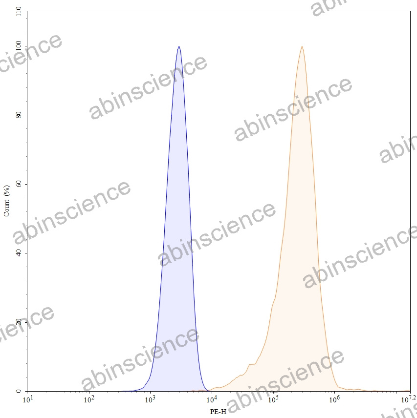 Flow CytoMetry |
Flow-cytometry using PE anti-human B2M antibody. Hela cells were stained with an irrelevant antibody (Blue Histogram) or an PE anti-human B2M monoclonal antibody (Catalog: HX250013, Yellow Histogram) at a concentration of 5 µg/ml for 30 mins at RT. After washing, and cells analysed on a NovoCyte Flow Cytometer. |